FALVEY LIBRARY

Seeing into “Triumph of David:” X-radiographs reveal hidden imagery

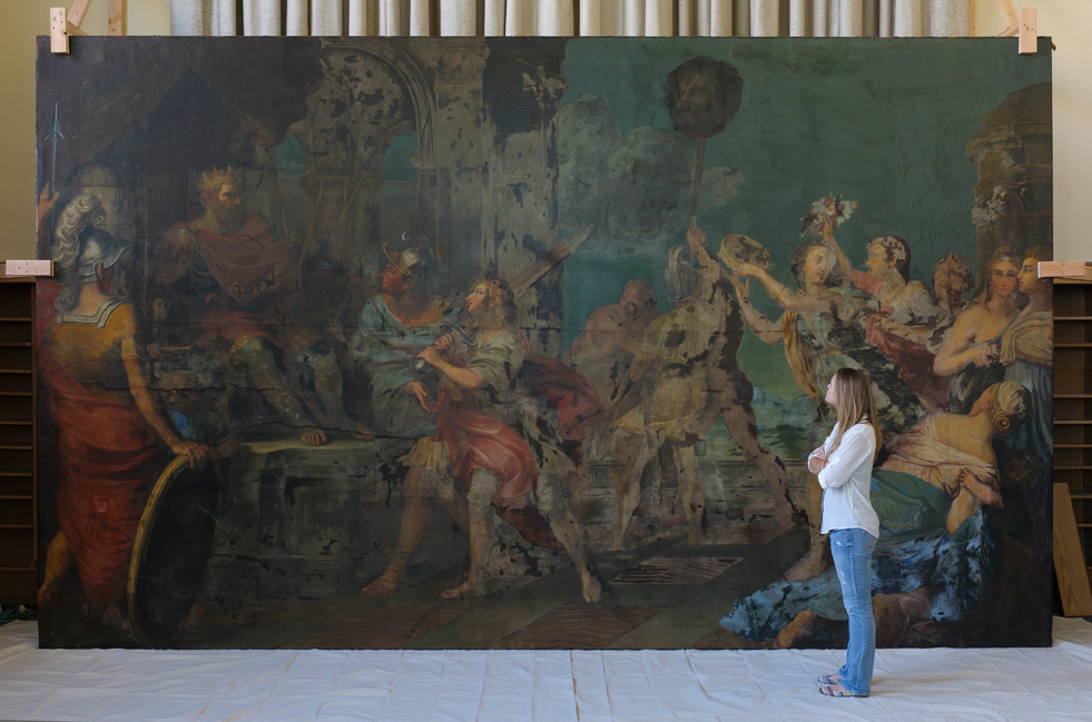
Just a little over two weeks ago, the Conserving a Giant conservation team was anxiously awaiting the arrival of technicians from General Electric (GE) Inspection Services Department, hoping to uncover secrets hidden under the paint using x-radiography.  With the generous support from the Samuel H. Kress Foundation, x-radiography was performed on Triumph of David and other paintings from the Villanova University Collection recently reunited in Old Falvey.

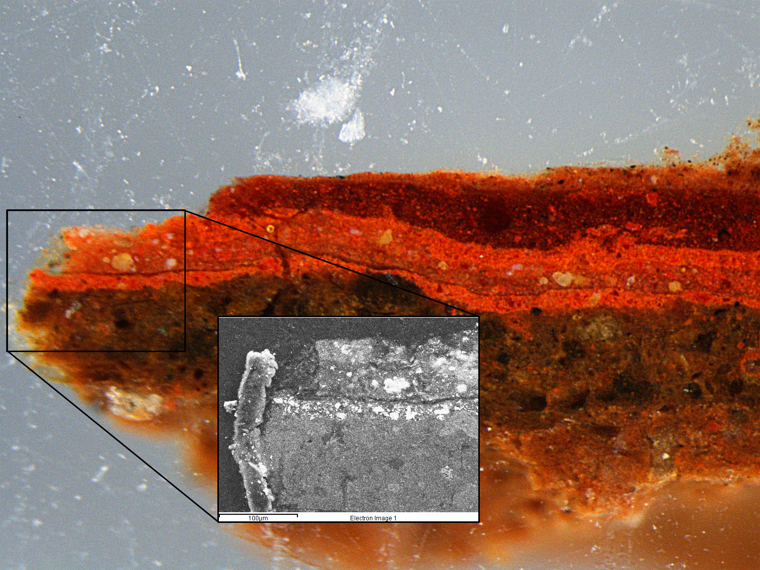
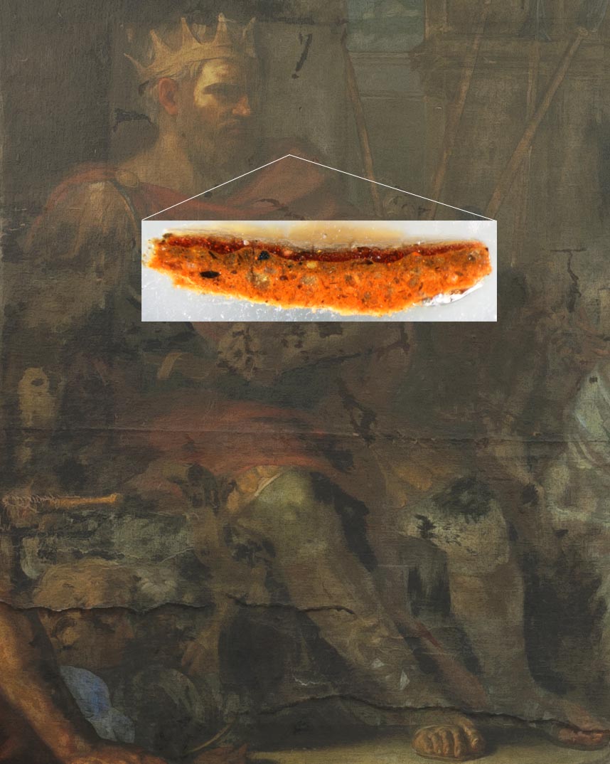
By directing x-rays through a painting and collecting the resulting image on radiographic film, x-rays can be used to highlight what is underneath the paint surface that we see today.  Some of the information that can be collected from the x-radiographs include pentimenti (compositional changes), areas of previous fills or older repairs, as well the location of tears and holes in the canvas or paint layers.

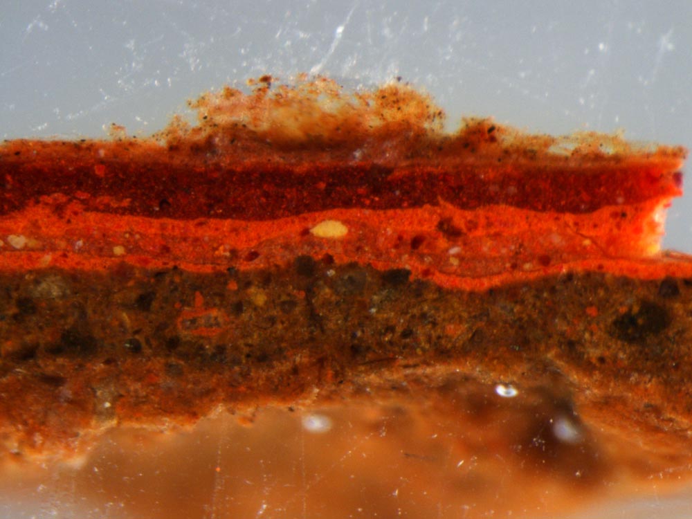
X-rays are shorter in wavelength than ultraviolet radiation, and the energy of that radiation allows the x-rays to penetrate through the paint surface.  The depth of penetration depends on the thickness and the density of the material that the x-rays are moving through.  For example, lower-density materials, such as the canvas or lighter weight elements such as carbon black, do not absorb as much x-ray radiation, and appear darker in the resulting x-radiograph.  Materials with higher densities, like heavy metal pigments such as cadmium red and lead white, absorb x-rays quite easily and show up as white or brighter grey in the x-radiograph.  One of areas that we were especially anxioius to learn more about was what might be located under the shield.  The shield is currently painted a flat black, and in comparison to the highly decorated regalia in the painting.  Conservators noted that there are brush stokes visible through the black paint that do not coincide with what is on the surface of the painting.  The chance to solve the mystery of what lies underneath those brushstrokes was eagerly anticipated.

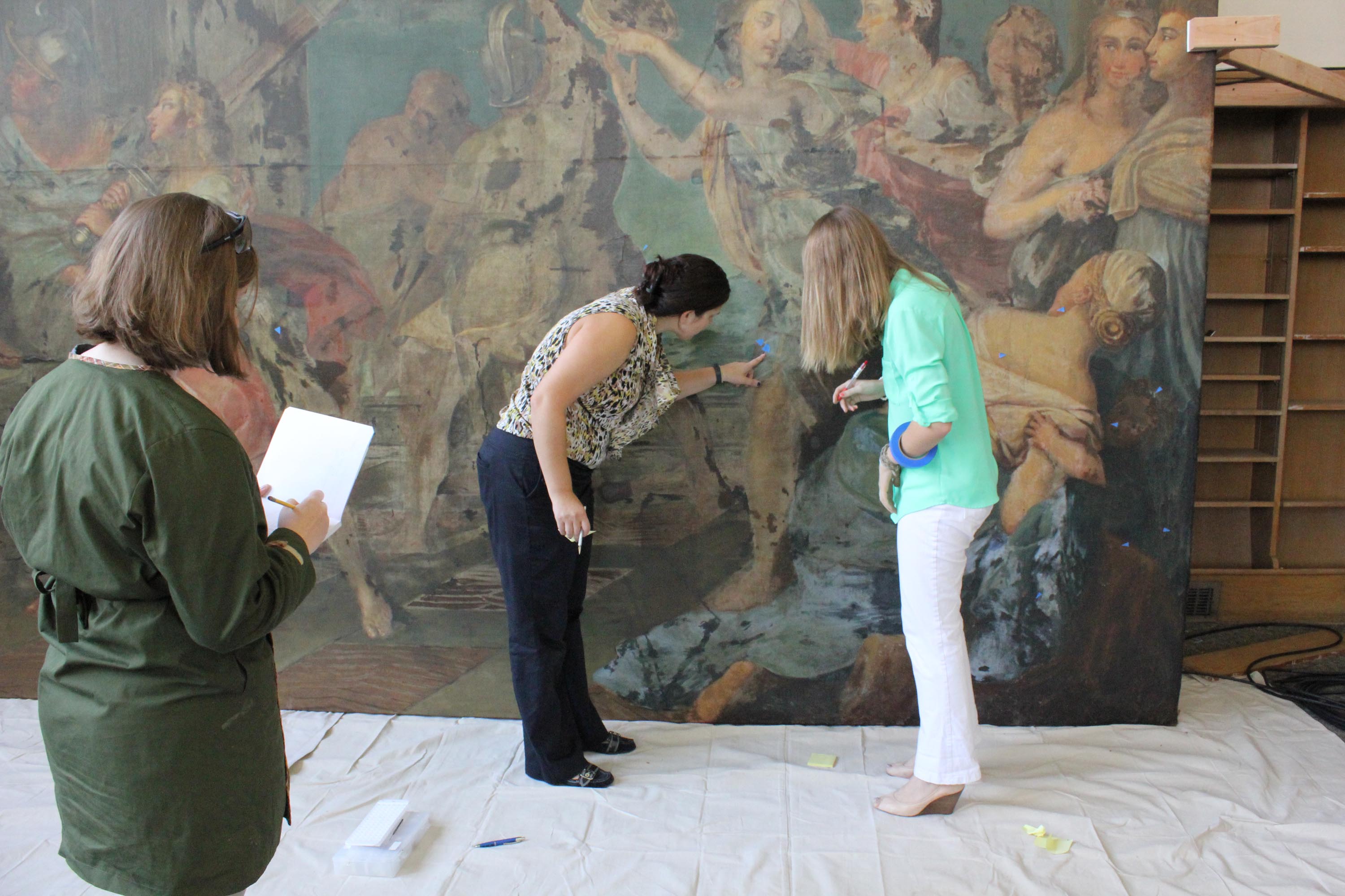
However, the conservation team first had to create a system for x-raying the painting, to be sure that areas of the painting would not be missed during the capture of over 100 x-ray images.  Another challenge facing the team was being able to complete the analyses of the large-scale painting in a two-day time span.  Prior to the arrival of the GE X-ray team, an alpha-numeric grid system was devised to aid in the positioning of the x-radiographs, ensuring an overlap of 1¾” between each exposure.

Slide1

Schematic of x-rays penetrating through
the painting, and being captured on the x-ray
film. The processed image is seen to the far right.

Dr. Anthony Lagalante and Brad Thorstensen from the Chemistry Department devised a novel method for securing the x-ray film to help maintain the grid system during data acquisition. This involved two steps; first, the location of each exposure was marked on the verso of the stretcher bars and second, a specialized film holder was designed to facilitate the process over a two-day time span.  A 22¾”x24½” x-ray film holder, designed by Thorstensen and Lagalante, was created using sheets of polycarbonate with rare-earth (neodymium) magnets mounted in each corner.  The magnets were padded with a soft synthetic rubber to protect the surface of the painting and an insert was created using 3M VHB tape.  Lines were also taped on to the holder as a guide for positioning the holder in the correct grid pattern. While this system worked well for The Triumph of David, it should be noted the painting is without impasto or cupping which afforded safe contact between the film holder and the painted surface.

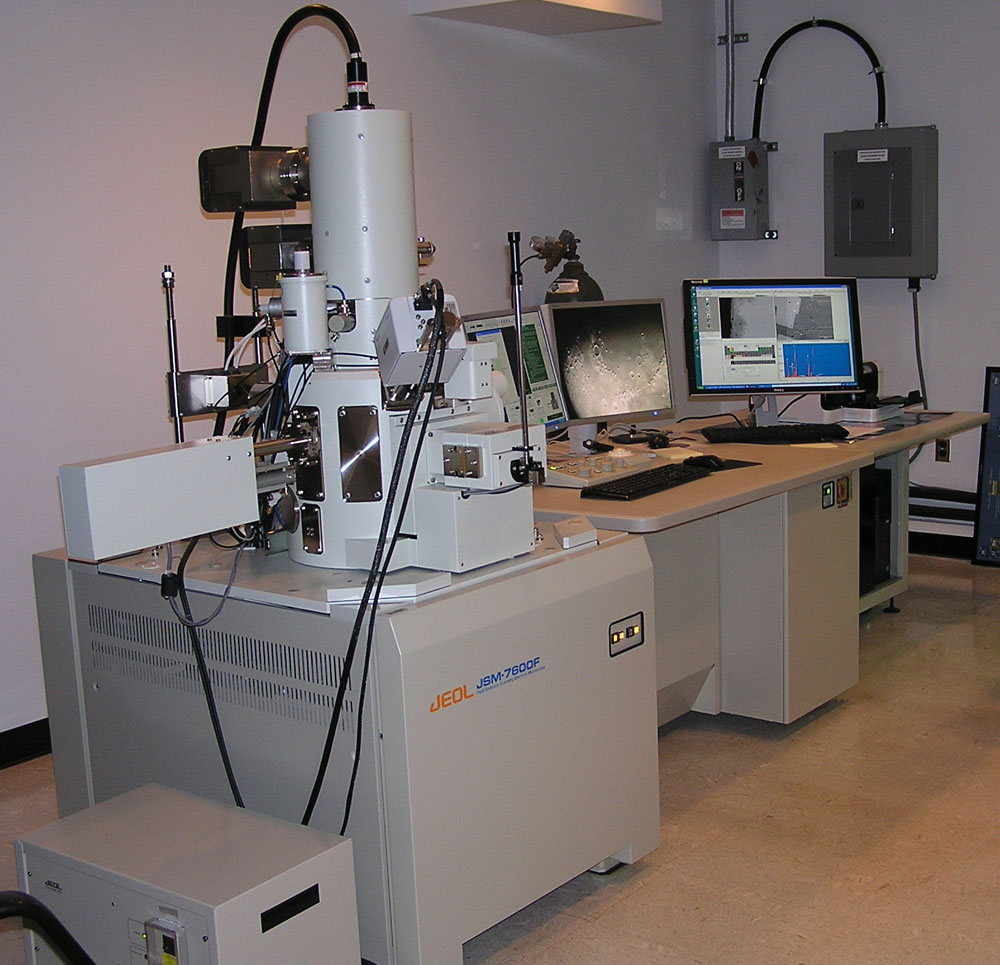
For the x-radiographic examination, the GE technicians used a Spellman LORAD LPX160 tube in conjunction with high-resolution 14″x17″ digital-based phosphor x-ray films. Images were captured at 30kV of radiation, which offered better quality in the resulting image.  To avoid any radiation exposure , the GE and Villanova teams stayed outside of the cage during capture times.  To be confident in safety, we also had ND-2000 dosimeters provided by James and Mark to measure the levels of radiation in the room throughout the imaging process.  (A safe level of radiation is 2 millrem per hour or less).   A GE Pegasus CR 50P digital scanner was moved into the space to scan the films as soon as they were exposed.  This allowed for immediate scanning of the films, meaning that we could readily view captured x-radiographs, and that multiple sheets could be used simultaneously.

LiveFeed

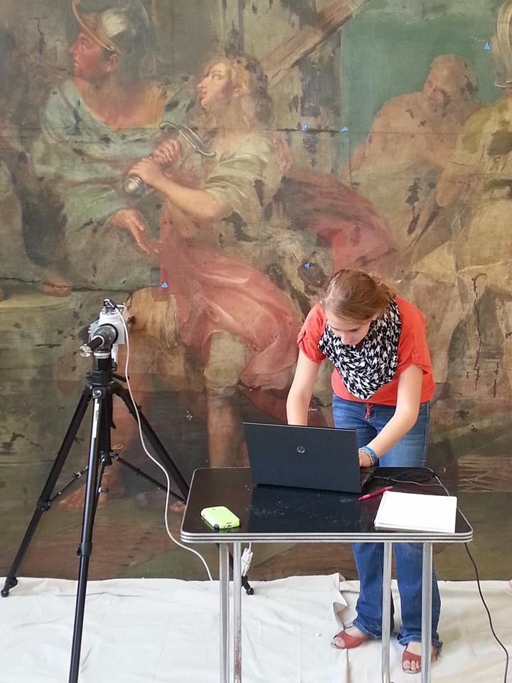
Kristin and Sarah helps align the front guide plate, while
Keara looks on. The green x-ray source is focused onto the
white x using a laser beam. The phosphor film is located just
behind the painting in the same location as the front guide.

Due to the sensitivity of the process, the Reading Room was closed off for the three days of setting up and x-raying Triumph of David and the other paintings in the collection.  However, remote access to the Live Feed allowed the general public to follow along as we made each capture across the entire surface of the painting.

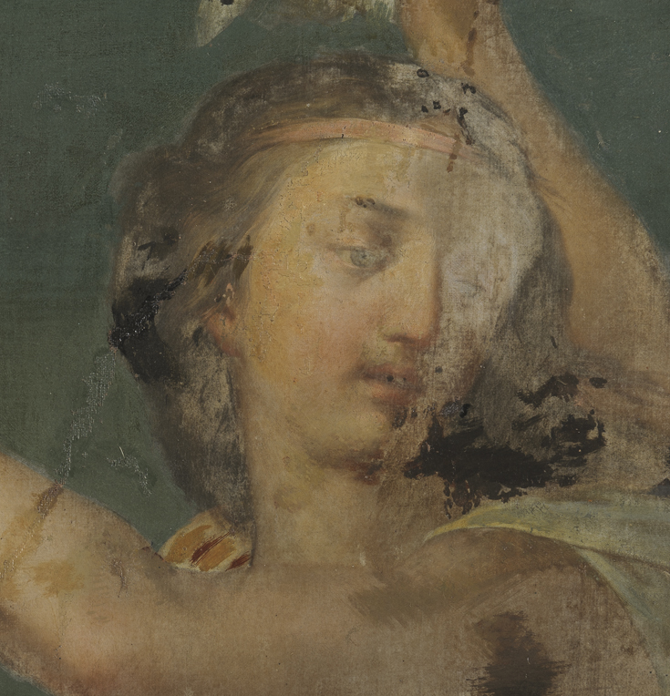
At the end of the first day, some of the captured x-radiographs provided insight in to the moving of David’s hands on the sword, and even the indecisiveness in the placement of Abner’s thumb.  The second day was spent reviewing the images and recapturing films that had too much excitation (“blown out” with too much white) or too little excitation (too dark).

But the greatest discovery was found in the paint layers beneath the dark shield.  In reviewing the captured images, a strange hand was seen holding some rod-like sticks, an object that was eventually determined to be a fasces.  Fasces are bound bundles of wooden rods equipped with an axe blade and were often associated with magisterial power in ancient Rome (there is even a fasces on the US dime).  Currently the team is faced with a new question: why was this man initially included in the composition and, more importantly, why was he painted out?

Figure in shield x-ray

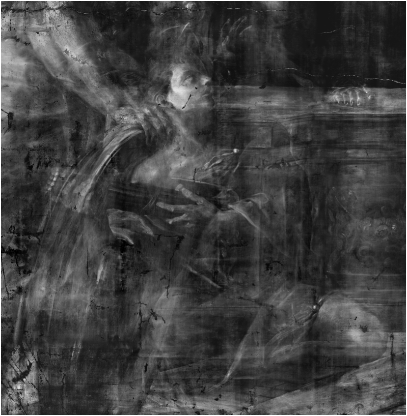
X-radiograph composite image. The figure is holding a
fasces, and the axe head can be seen just above the left
hand of the hidden man.

Sheild area

How the sheild area appears today.


In the end, over 120 images were captured, which lead conservator Kristin deGhetaldi compiled into the final image seen below. To learn more, please come visit Old Falvey, as we are very excited to share the discoveries made while examining Triumph and the other paintings in the Ruspoli collection with x-radiography!

Pietro During      Composite

We will leave you with the composite image of all of the x-radiographs taken of the Triumph of David. Looks amazing. Leave a comment below if you can see any differences between the painting and its x-radiograph!!

Generous Support from the Samuel H. Kress Foundation Allows Conservation Team to X-Ray “Triumph of David”

We are most pleased to announce that, in May 2014, the project Conserving a Giant: Resurrecting Pietro da Cortona’s “Triumph of David” was awarded a substantial grant from the Samuel H. Kress Foundation. These funds will be utilized in a number of ways, to help both produce and disseminate further information knowledge about Villanova University’s large canvas – and no doubt about seventeenth-century Italian painting more broadly – to in a number of arenas and in a variety of audiences, both general and academic. 

The Kress foundation praised the interdisciplinary nature of the project, which has involved thus far a number of department and offices at Villanova, and the participation of scholars and students of art conservation, chemistry, history, and art history. We are very happy that this support will be used to further specifically interdisciplinary investigations, and that it will continue to allow Villanova faculty to engage this painting in both teaching and research, with both undergraduate and graduate students, and with both the public at large and the wider academic community.  This grant will allow us to involve even more students, professors, and departments on campus, and it will enable us to develop further cooperation with art historians of Roman Baroque painting, art conservators, and scholars at institutions with connections to the painting and its donor. 

Most immediately, the Kress funding will allow for technicians from General Electric to perform X-radiography and other technical analysis on the gigantic canvas, and other early modern paintings in Villanova’s collection. Several large canvas paintings by Pietro da Cortona were examined using X-radiography during a technical study in 1997-8, revealing characteristic unique to the artist’s working method.  X-radiography of the Villanova painting will allow the conservation to establish a dialogue with other scholars and art historians who are more familiar with Pietro da Cortona as well as artists related to his circle.

With this grant, members of the Conserving a Giant conservation team and Falvey Library will collaborate with Villanova University’s Computing Sciences Department and with UNIT (the IT Department) to create a “webexhibit” exploring the Triumph of David. This website will enable members of the conservation team as well as chemistry and art history faculty and students to compile, organize, and share their research with a wider audience. The “webexhibit” will remain a permanent fixture on Villanova’s server, thus providing an attractive and interactive site for prospective students, outside scholars, and the general public, and moreover, serving chemistry and art history courses for years to come.

Funding from the Samuel H. Kress Foundation will allow members of the team at Villanova to travel to Rome in spring of 2015, to view a number of frescoes and canvas paintings by the artist, and by important members of his workshop, to gain further insights about his painting methods.  We will be meeting with art historians and conservators who have worked on seventeenth-century painting, to learn from them and, with what we have discovered through conservation and research undertaken at Villanova, to share our own experiences and knowledge. The grant, together with support from Villanova University, will additionally fund an international, interdisciplinary symposium investigating Pietro da Cortona’s workshop in seventeenth-century Rome. This symposium will involve a number of scholars and students from various fields who have contributed to the Conserving a Giant project, and we will be able to invite eminent scholars of Baroque painting. The symposium will serve as both a culmination to the multi-year conservation project, and at the same time a new beginning, with new knowledge and insights shared among and between established and emerging art historians, scientists, and conservators, and, indeed, with all of the Villanova community.    


– Tim McCall

Professor of Art History

Villanova University


Pigment Discovery with Electron Microscopy

  • Posted by: Kristen Watts
  • Posted Date: March 6, 2014
  • Filed Under: Chemistry

As we (slowly) move towards spring, the science team has been working on the microsample cross-sections from Triumph of David using our suite of analytical instrumentation.  As we noted in our previous entry, if a sample is going to be removed from a painting, we are going to try to get the most amount of information from it as possible. To investigate questions of pigmentation beyond the color profiles seen with bright-field microscopy, we move on to a new technique called scanning electron microscopy paired with energy-dispersive x-ray spectroscopy (SEM/EDS).


Photo of an SEM

Photo of a SEM

Harkening back to the science blog post describing how we use fluorescent X-rays to noninvasively image and characterize the painting, SEM/EDS utilizes a similar method to analyze pigments in our cross-sections. XRF is useful in giving us a general idea of the elemental breakdown of inorganic pigments in an area of the painting; however, because of the high penetration power of the x-rays, and the fact that it is a non-invasive characterization method, we have no way of telling whether the elements we are finding in an area is a part of the ground layer, pentimenti, or a restoration campaign. SEM is a form of microscopy in which the sample is bombarded by a high energy electron beam, which can give very precise images (up to 50,000 times magnification!) based on the collection of the scattered electrons that have bounced off the surface. We can then use these images to collect elemental maps of the layers using EDS software. EDS detects fluorescent x-rays, just like XRF instruments, but with much higher spatial precision.


Location of an SEM image on the overall cross-section

Location of an SEM image on the overall cross-section

The particular sample we’re discussing today was mentioned at the end of the last science blog post, taken from the red cloak of the heavily overpainted soldier on the left-hand side of the painting (see image to the left). With the enhanced magnification capability of the SEM we can get precise information concerning the inorganic materials down to the individual pigment particles. This particular SEM image, the black and white image in the photo to the left, was taken at 450x magnification, and provides a wealth of information concerning the sample.


In the main SEM image, it’s pretty clear to see that there’s at least 5 layers of paint in this one subsection. By combining this SEM image with elemental mapping data, we can see the general elemental breakdown for each of these layers. The bottom layer, with most of the calcium, silicon, potassium, and aluminum is probably some sort of clay-based ground, as seen in the image below.

Comparison of the Silicon (Si) elemental map with the main SEM image

Comparison of the Silicon (Si) elemental map with SEM/BSE image

There then appears to be a layer of vermilion (as evidenced by the presence of mercury and sulfur–HgS) before a thin layer of organic material (the thin dark layer in the SEM image) was applied, shown below.

Comparison of Mercury (Hg) elemental map with SEM/BSE image

Comparison of Mercury (Hg) elemental map with SEM/BSE image

The layers above the organic layer are where the cross-section becomes incredibly more complex, including a mixture of vermilion, aluminum-based lakes, lead white, and even some Naples yellow, as seen by the spectral evidence below.


An example EDS spectrum showing the presence of lead (Pb) and antimony (Sb) in one particle--indicating the presence of Naples yellow

An example EDS spectrum showing the presence of lead (Pb) and antimony (Sb) in one particle–indicating the presence of Naples yellow

As you can see, there’s a lot of information to be gleaned from such a small sample, and there is still a lot that is unknown and up for interpretation. But we’re analyzing everything piece by piece and can hopefully provide more answers in the future. Until next time!


Peeling Back the Layers

  • Posted by: Kristen Watts
  • Posted Date: December 6, 2013
  • Filed Under: Chemistry
Amanda Norbutus and Kristen Watts discuss sampling locations with Emily Wrozenski taking notes.

Amanda and Kristen discuss sampling locations with Emily taking notes.

The chemistry team has been busy this past month sampling and preparing cross-sections for analysis from The Triumph of David. As mentioned last time, x-ray fluorescence (XRF) is a technique that allows us to get a broad overview of what inorganic elements are present in a particular area of a painting. Usually, XRF can be  incredibly useful in getting an idea of what pigments may be present in the area being analyzed. However, since the x-rays can penetrate all the way through to the canvas of the painting, we have no way to tell in which paint layer the detected elements are present by XRF alone. In order to determine which layer the pigment is located, we need to take a sample and prepare a cross-section. Cross-sections are beneficial for our research in that they not only allow us to visualize the different layers of paint present in an area, but also, with new developments in microscopy, we can determine which inorganic materials and binding materials are present in each layer as well.

Example of a cross section taken from King Saul's red cloak.

Example of a cross section taken from King Saul’s red cloak.

Sampling  from a 17th century painting isn’t quite as scary or taboo as it might seem at first. While the removal of anything original to the painting is not ideal, sometimes a sample is necessary in order to get the most accurate idea of the make-up of the painting. Conservators and scientists will take micro-samples, often no bigger than the period at the end of this sentence.  We micro-sample from areas of pre-existing loss: meaning areas where the paint has already started to flake away. In the case of The Triumph of David, there were innumerable areas of loss from which we could sample. Obviously, we couldn’t and didn’t want to sample from every area of loss; so, we chose our sampling locations partially based on where the XRF gave intriguing results and partially in areas of interest to the conservators–namely, areas that appeared to be heavily overpainted or areas where it was difficult to distinguish pentimenti from later restoration campaigns. The micro-sample is then embedded in a plastic and cut and polished until the cross-section is exposed as in the figure above.  This single exposed cross-section can be examined using a vast array of microscopic techniques which will be expounded upon in future blogs.

Sampling location of aforementioned cross section

Sampling location of aforementioned cross section

This sample in particular was taken from the red cloak on King Saul, and the image above was taken at 80x magnification. We took a sample in this location because we were curious to see if Saul’s cloak had been repainted and also to see if the red pigments matched other red pigments seen throughout the painting. As shown in the full size cross section above, there are obviously very different paint layers present in this one cross-section. The bottom two tan/brown layers are called ground layers. A ground layer is present in any painting and serves as a preparatory layer, creating a uniform surface on which to paint. Layer 1 appears to be made with less finely ground particles than layer 2, as you can see from the bigger particles present in layer 1. This means that layer 1 provided an initial ground layer to size the canvas, whereas the second layer created a uniform paint surface. Layer 3 is the first intentionally pigmented red paint layer that provides the coloring for Saul’s cloak. Finally, Layer 4 is the 1956 re-varnish present on the entire painting that the conservators are actively removing. Varnish serves the purpose of bringing more saturation to a painting as well as providing protection for the paint underneath. It is this varnish that has degraded over the years and has accumulated dirt which can be seen in the cross-section.


032-11

Cross section taken from the left side soldier’s overpainted red cloak.

However, not all cross sections are this straight forward to interpret. A good example is the cross-section, shown to the right, taken from a soldier’s red cloak, an area that may have been re-painted more than once during previous restoration campaigns. Cross sections, especially with a painting of this age and indefinite history, can be incredibly complex and often yield more questions than answers. Frequently, it becomes difficult to tell which layers are original and which are from subsequent restoration attempts. Cross sections like the one above require more than just a visible light micrograph in order to fully interpret the image. Techniques that go beyond what our eyes are capable of seeing, such as fluorescence microscopy and scanning electron microscopy, are incredibly useful as they can help us understand the binding materials and inorganic pigments used in each layer. So stay tuned as we keep unraveling the mystery of Pietro da Cortona’s The Triumph of David.


Until then, Ciao!

-Kristen, Amanda, and Anthony.


ZAP! Visualizing Pigments with X-Rays

  • Posted by: Kristen Watts
  • Posted Date: October 30, 2013
  • Filed Under: Chemistry
Kristen Watts working on XRF analysis of Triumph of David

Kristen Watts using XRF to analyze The Triumph of David

As The Triumph of David conservation project gains momentum, we, the chemistry team, have been studying the materials used in the painting. Conservation science is considered science in the service of cultural heritage and is a field of study that most science majors don’t even know exists.  Conservation scientists are interested in the chemistry of artistic materials and techniques and how they age in an artwork’s environment.   The proliferation of high-powered analytical instrumentation has made it possible to analyze historical objects with in-depth, non-destructive techniques. Our role in the conservation team is to provide data to conservators that can identify the materials used by the original artists as well as the materials used during subsequent restoration campaigns (a campaign refers to restoration efforts separated by time).  We are also hoping to provide the art historians with technical information that may help with dating and understanding how Pietro da Cortona and his workshop painted this rare, large format, oil-on-canvas painting.  Ultimately, we would like to provide a scientific answer as to why the varnish and the areas of obvious overpaint have darkened and deteriorated so quickly since the most recent restoration campaign in 1956. At this moment, we are busy trying to identify the pigments present in the painting as well as which paint layers are original and which are restoration.

Inorganic pigments, like verdigris or vermillion, can often be identified utilizing x-ray fluorescence (XRF) and scanning electron microscopy paired with energy dispersive x-ray analysis (SEM-EDX). During the past few weeks, we have been focusing our work on XRF as well as optical microscopy of cross-sections that have been taken, but today we are going to focus on some of our XRF results.

Schematic of XRF process

XRF is incredibly useful in conservation science not only because it’s nondestructive but also because portable, hand-held instruments are available, meaning that the instrument can be brought to the painting instead of bringing the painting to the lab where the instrument resides. XRF utilizes high energy X-rays to eject inner shell electrons from an element. The outer shell electrons will then fill the electron hole, releasing an x-ray photon in the process. This photon release is known as fluorescence and the particular energy of the released photon is specific to each element. Because of this specificity, we can use XRF to help us identify inorganic pigments in areas of interest. (For a more in depth look at the XRF mechanism of action, see here)


XRF spectra of the yellow shirt and the red toga on the soldier.

XRF spectra of the yellow shirt and the red robe on the soldier.

In the case of the soldier’s red robe (as seen in the photo at the left) we found a signal for mercury (Hg), indicating that the red color is probably due to a vermillion (mercury sulfide) pigment. There was also a large signal for lead (Pb) but this signal is found throughout the painting as it is caused by a lead white containing ground layer. His yellow shirt, on the other hand, showed signs of chromium(Cr) and barium (Ba) which would indicate a more modern chrome yellow (post 1800) or lemon yellow (post 1830) paint, thereby indicating that one of the paint layers originates from a more modern restoration campaign.


As mentioned previously in the Metigo map blog post, there are a lot of areas with heavy overpaint present as well as several restoration campaigns. By using XRF to monitor the presence of zinc (Zn), we can assist the conservators in tracking the removal of the restoration materials. Currently, zinc is suspected to be a component of the varnish or in a slightly pigmented glaze from a previous restoration. We suspect this because we see zinc disappear significantly with varnish removal, as seen in the image below, comparing the uncleaned overpaint (red), cleaned but still overpaint (green), and cleaned without overpaint (pink) all on a soldier’s leg in the painting. The spectrum corresponding to the uncleaned overpaint shows a moderate zinc signal, whereas zinc is not detected in the spectrum corresponding to an area of overpaint with just the varnish removed and the spectrum corresponding to a completely cleaned area.


Comparison of uncleaned overpaint, cleaned overpaint, and totally cleaned areas present in a soldier's leg

Comparison of uncleaned overpaint (red line), cleaned overpaint (green line), and totally cleaned (pink line) areas present in a soldier’s leg


Because XRF simultaneously examines all the layers of the painting from the ground, paint layers, over-painted layers, and varnish, we can’t tell exactly in which layer the pigment is present by XRF alone. However, this XRF data is useful when determining where to micro-sample the painting to get layer-specific pigment information through techniques such as SEM-EDX and fluorescence microscopy, which you can read about in later posts. We are just beginning to unravel some of the mysteries surrounding this painting and our team is learning more every day.

Until next time, Ciao!

Anthony, Amanda, and Kristen.

Conserving a Giant: Resurrecting Pietro da Cortona’s “Triumph of David”

da-Cortona-1

Welcome to our weekly blog detailing the conservation treatment of Pietro da Cortona’s “Triumph of David”, a large-scale oil on canvas that currently resides in “Old Falvey,” Falvey Memorial Library’s original wing. The painting first came to the University in 1956, donated by the late Princess Eugenia Ruspoli who inherited the artwork from her late Italian husband, Prince Enrico Ruspoli. For much of its life, the “Triumph of David” hung within the walls of Castle Nemi. The castle sustained significant damages during World War II which may partly explain the current condition of the painting. Pietro Berrettini da Cotrona was perhaps best known for his work as an architect, however the artist and his workshop executed several exquisite fresco cycles in addition to a number of canvas paintings. As only a hand-full of Pietro’s canvas paintings can be found in museum collections, the University is make a concerted effort to restore the artwork and promote collaborative research amongst art historians, conservators, and scientists. More information can be found in the recent press release.

Villanova now joins a hand-full of cultural institutions and conservation laboratories that have begun to embrace the concept of “visible conservation.” A permanent space in Old Falvey’s reading room has been allocated for the project, allowing students as well as the general public to observe the treatment process and even interact with the conservation team. A schedule will soon be posted, listing dates/times for formal tours and a live web-cam will add an interactive element to the project. This will help to raise awareness of contemporary conservation methods, the history of the painting, and the upcoming renovation of the Library’s reading room.

Collaboration on Campus

An exciting aspect of the project will involve the collaborative efforts of scholars, faculty, staff members, and students, interactions that will occur on a daily basis in the publically accessible conservation space. Dr. Anthony Lagalante, Dr. Amanda Norbutus, and graduate student Kristen Watts from the Chemistry Department will be working alongside the conservation team, performing pigment analysis in an effort to learn more about the materials used by Pietro and his workshop. Dr. Mark Sullivan, Dr. Timothy McCall, and senior undergraduate student Rachel Godat from Art History will be offering their expertise as well, helping the conservation team to gather more information relating to the artist, his workshop, and other aspects of the painting’s provenance. Finally, Darren Poley and the library staff will be integral in the promotion of the project and it’s connection to the upcoming renovation of the Library’s Old Wing. Future events will be announced as the treatment progress and will likely include lectures, symposia, web projects, and/or interactive workshops.

The Conservation Treatment

Detail Showing OverpaintThe treatment of the painting will be carried by painting conservator Kristin deGhetaldi over a period of two years with the help of conservation interns and graduate students (currently Maggie Bearden and Emily Wroczynski). The painting is presently covered with several layers of discolored and degraded over-paint and varnish, unoriginal materials that have aged poorly and now obscure the original colors of Pietro’s vibrant palette. Conservators are professionals who have specialized knowledge and skills in the arts, sciences, and other fields; responsibilities can include establishing appropriate environmental controls, stabilizing the structure of an object, compensating for surface disfigurement from deterioration and/or damage, and undertaking technical studies. A professional conservator conducts these activities according to the Code of Ethics of the American Institute for Conservation of Historic and Artistic Works.

In years past, those interested in becoming a conservator would typically enlist as an apprentice in a restoration studio. Today, however, most students pursue a master’s degree in art conservation after obtaining a bachelors degree in art history, studio art, the sciences, or other related fields. In order to be accepted into a graduate degree program in the United States, students must satisfy extensive course requirements (e.g. organic chemistry, art history/anthropology courses, a studio art portfolio, etc.) and complete a certain number of hours serving as a volunteer/intern in a cultural institution or private studio. This pre-program experience often takes additional time beyond completion of an undergraduate degree. Students who are interested in learning more about graduate programs should also visit the Emerging Conservation Professionals Network blog.


Last Modified: October 11, 2013